Los ganadores del concurso olympus Bioscapes, realmente espectaculares (sigue leyendo para ver el resto)
Olympus BioScapes Competition Winners (9 Images)
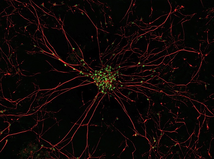
© Gist Croft and Mackenzie Weygandt, Columbia University and Project ALS, New York, N.Y.
Amyotrophic Lateral Sclerosis (ALS, also known as Lou Gehrig’s Disease) motor neurons. The stem cells used to generate these motor neurons were induced Pluripotent Stem (iPS) cells made from the skin cells of an 83-year-old ALS patient.
This image won tenth prize in the Olympus BioScapes Digital Imaging Comptition, an international competition of microscope imagery. The winners were announced November 18, 2009. This year, the competition received nearly 2,000 entries from 62 countries. All images and the names of the top winners and Honorable Mentions may be viewed online, and some of images will be part of a traveling exhibition.
A selection of winning images follows.
© Haruka Fujimaki, Bryant Pond, Maine
Atlantic salmon embryos. Ninth prize.
© Heiti Paves, Tallinn, EstoniaFlower of Arabidopsis thaliana (thale cress), a popular model organism in plant biology and genetics. Image captured using confocal microscopy using a 20x objective lens. Eighth prize.
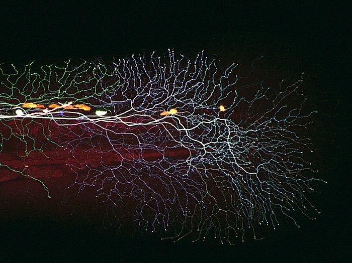
© Albert Pan, Harvard University, Cambridge, Mass.
Sensory axons (long, slender nerve fibers) covering the tail of a 3-day-old larval zebrafish. This is a “Brainbow” image made using confocal microscopy. In the Brainbow technique, cells randomly choose combinations of red, yellow and cyan fluorescent proteins, so that they each glow a particular color. This provides a way to distinguish neighboring cells of the nervous system and follow their pathways. Seventh prize.
© Alvaro Migotto, University of São Paulo, São Paulo, BrazilTentacle of Portuguese Man o’ War, Physalia physalis, magnified 30x. Notorious for its painful, powerful sting, the Portuguese Man o’ War has a gas-filled floating chamber that supports the tentacles, which bear sting cells. Shown are the pink batteries of stinging cells and a delicate muscular band responsible for the high contractibility of the tentacles. Sixth prize.
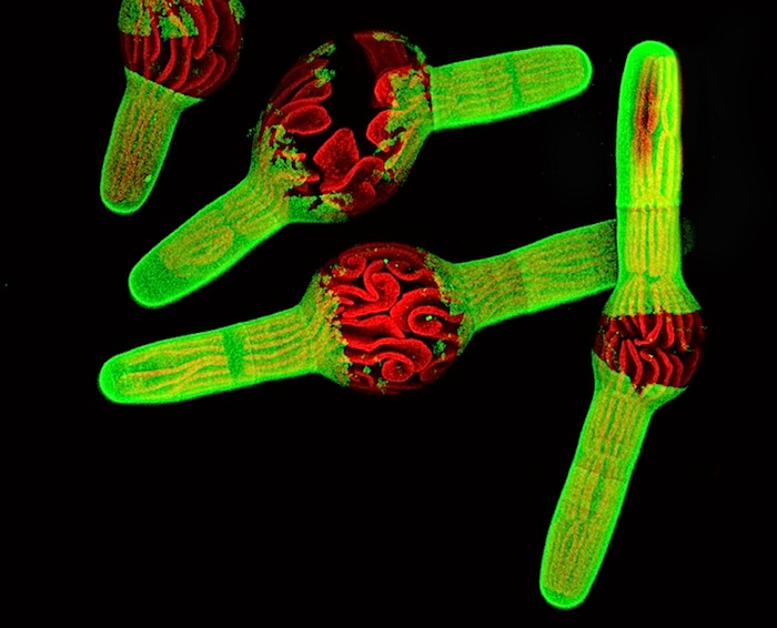
© David Domozych, Department of Biology, Skidmore College, Saratoga Springs, N.Y.
Unicellular alga Penium, treated with the microtubule poison oryzalin. Fifth prize.
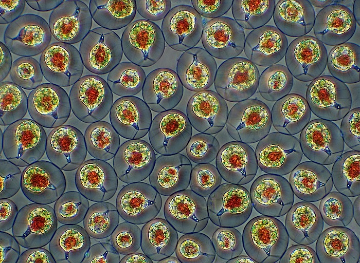
© Charles Krebs, Issaquah, Wash.
Fresh water algae Haematococcus pluvialis, 100x. Phase contrast microscopy. Fourth prize.
(Third prize went to a time-lapse video of algae cells by Jeremy Pickett-Heaps of the University of Melbourne, Australia.)
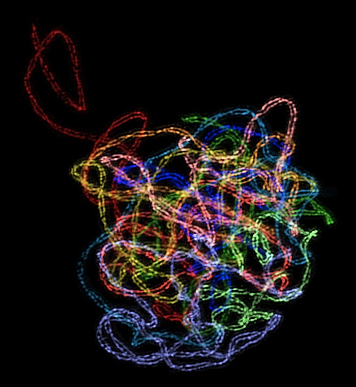
© Chung-Ju Rachel Wang, Department of Molecular and Cell Biology, University of California, BerkeleyNucleus of a plant cell showing synaptonemal complex, a ladder-like protein structure that forms between pairing chromosomes
during meiosis (the cell division required for reproduction). This may be the first-ever high-resolution 3D image of this complex
ever captured with light microscopy. The two parallel axes of this complex, which run the length of each chromosome, are seen as two threads spaced 100-200 nm apart and twisting around each other in a helix. Second prize.
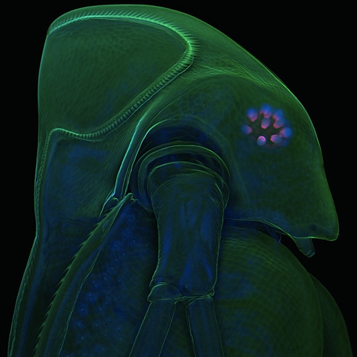
© Jan Michels, Department of Functional Morphology and Biomechanics, Institute of Zoology, Christian Albrecht University of Kiel, Germany.Water flea Daphnia atkinsoni. This specimen has a “crown of thorns,” a defensive trait induced in offspring only when the parents sense chemical cues released by one of their main predators, the tadpole shrimp Triops cancriformis. The water flea’s exoskeleton (exterior structure, green) and subcellular details within the organism (nuclei – tiny blue dots) are both visible. First prize.
Olympus BioScapes Competition Winners (9 Images) | PDN Photo of the Day

